Summary: A breakdown in regulatory mechanisms causes iron to build up in the brain during aging, increasing oxidative stress and increasing the risk of age-related cognitive decline, a new study reports.
Source: Northwestern University
Breakdowns in regulatory mechanisms cause iron to build up in the brain as organisms grow older, increasing oxidative stress and causing cellular damage, according to a Northwestern Medicine study published in the journal eLife.
This mechanism may explain some age-related cognitive decline and contribute to neurodegenerative diseases such as Parkinson’s and Alzheimer’s disease, according to Hossein Ardehali, MD, Ph.D., the Thomas D. Spies Professor of Cardiac Metabolism and senior author of the study.
“There is tight regulation of iron homeostasis in the brain, but it appears that this regulation is disrupted as we age,” said Ardehali, who is also director of the Center for Molecular Cardiology at the Feinberg Cardiovascular and Renal Research Institute. “There are studies planning to use iron chelators in coronary artery disease and exploring these in the brain and aging is the next step.”
As organisms age, oxidative stress increases in cells across the body. For one reason or another, cells lose the ability to detoxify reactive oxygen species, byproducts of normal cellular respiration.
The source of oxidative stress varies from environment to environment within the body, but previous studies point to one possible source in the brain: an accumulation of iron, according to Ardehali, who is also a professor of Medicine in the Division of Cardiology and of Pharmacology, and a member of the Robert H. Lurie Comprehensive Cancer Center of Northwestern University.
In the current study, Ardehali, along with first author Tatsuya Sato, MD, Ph.D., assistant professor at the Sapporo Medical University School of Medicine and a former postdoctoral fellow in the Ardehali laboratory, examined young and aged mice, measuring both cytoplasmic and mitochondrial iron throughout the body.
The investigators found the brain was the only organ which showed an increase in both cytoplasmic and mitochondrial iron as the animals aged.
Next, Sato examined expression of genes associated with iron homeostasis, finding that the gene coding for a peptide hormone—hepcidin—was dramatically upregulated in the brain cortex of older animals.
Hepcidin is a hormone produced by the liver that controls systemic iron homeostasis, but in the context of this study, brain-derived hepcidin’s most important function is inhibition of ferroportin, a protein that exports iron from the neuronal cells, leading to marked iron accumulation in the aged brain.
“This is likely a key player in iron accumulation in the aged brain,” Sato said.

The detailed mechanism of increased brain-derived hepcidin expression in the aged brain requires further study, but age-related inflammation and increased expression of iron-sensing protein transferrin receptor 2 may be possible regulators, Ardehali said. Increased hepcidin also may lead to increased iron in the mitochondria—more than the organelles can utilize—leading to buildup of iron and eventual cell damage. This finding spotlights a possible therapeutic strategy, according to Sato.
“If we can restore intracellular iron levels via suppressing this brain-derived hepcidin, we might be able to improve age-related cognitive decline,” Sato said.
There are ongoing studies using iron chelators—substances that bind to iron and make it biologically unavailable—to treat coronary artery disease, and Ardehali said a similar strategy could be used in the brain, as well.
The wrinkle is in the specific compound: to reduce iron concentration in the brain would require a chelator that can pass the blood-brain barrier. However, there is an ongoing clinical trial assessing the effects of a specific iron chelator in Parkinson’s disease.
“Not all chelators get though the barrier but one does. Exploring this possible therapy is our next step,” Ardehali said.
About this aging and cognition research news
Author: Will Doss
Source: Northwestern University
Contact: Will Doss – Northwestern University
Image: The image is credited to Northwestern University
Original Research: Open access.
“Aging is associated with increased brain iron through cortex-derived hepcidin expression” by Tatsuya Sato et al. eLife
Abstract
See also

Aging is associated with increased brain iron through cortex-derived hepcidin expression
Iron is an essential molecule for biological processes, but its accumulation can lead to oxidative stress and cellular death. Due to its oxidative effects, iron accumulation is implicated in the process of aging and neurodegenerative diseases. However, the mechanism for this increase in iron with aging, and whether this increase is localized to specific cellular compartment(s), are not known.
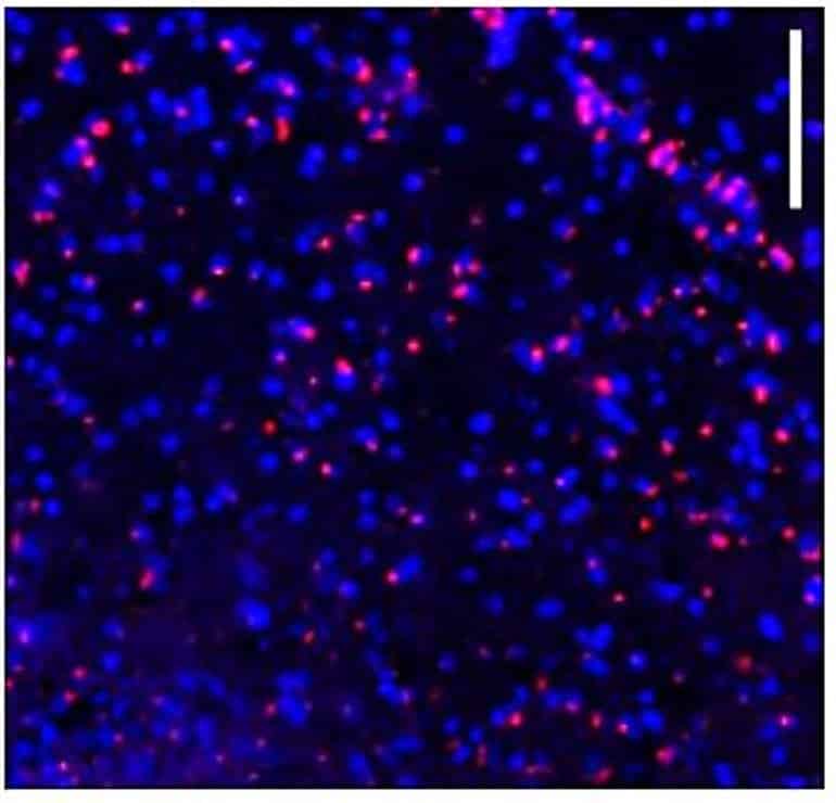
Here, we measured the levels of iron in different tissues of aged mice, and demonstrated that while cytosolic non-heme iron is increased in the liver and muscle tissue, only the aged brain cortex exhibits an increase in both the cytosolic and mitochondrial non-heme iron. This increase in brain iron is associated with elevated levels of local hepcidin mRNA and protein in the brain.
We also demonstrate that the increase in hepcidin is associated with increased ubiquitination and reduced levels of the only iron exporter, ferroportin-1 (FPN1). Overall, our studies provide a potential mechanism for iron accumulation in the brain through increased local expression of hepcidin, and subsequent iron accumulation due to decreased iron export.
Additionally, our data support that aging is associated with mitochondrial and cytosolic iron accumulation only in the brain and not in other tissues.
Credit: Source link


